Tagged: Priscilla Corraini
Periodontal Myths and Mysteries Series (I): How to Measure Attachment Loss?
As many readers may have noted [1], I had been quite concerned about how clinical attachment level (CAL) had been measured in the 2009-2010 continuous NHANES which had been reported in 2012 by Eke et al. (Note: 2011-2012 continuous NHANES has been completed and periodontal results are probably being published soon). After having adopted full-mouth, including 6 sites per tooth, recording and a different case definition, authors had reported quite dramatic higher prevalence of, in particular, moderate periodontitis in the adult population of the U.S. as compared to prevalence reported in NHANES III (Albandar et al. 1999). Another possible amendment was how CAL was calculated.
As for NHANES III, Albandar et al. (1999) had explained the procedure as follows.
The distance from the cemento-enamel junction (CEJ) to the free gingival margin (FGM) and the distance from the FGM to the bottom of the pocket/sulcus were assessed at the mesio-buccal and mid-buccal surfaces. The measurements were made in millimeters and were rounded to the lowest whole millimeter. The assessment was made by using the NIDR periodontal probe. The probing depth was defined as the FGM/sulcus measurement. The CEJ/FGM distance was given a negative sign if the gingival margin was located on the root. Attachment loss was defined as the distance from CEJ to the bottom of the pocket/sulcus and was calculated as the difference between CEJ/FGM and FGM/sulcus distances (or the sum of the 2 distances if FGM was on the root).
Interestingly, Albandar et al. (1999) do not mention recession but rather distances between well-defined landmarks. In contrast, Eke et al. (2012) describe,
All periodontal examinations were conducted in a mobile examination center (MEC) by dental hygienists registered in at least one U.S. state. Gingival recession [= the distance between the free gingival margin (FGM) and the cemento-enamel junction (CEJ)] and pocket depth (PD) (= the distance from FGM to the bottom of the sulcus or periodontal pocket) were measured at 6 sites per tooth (mesio-, mid-, and disto-buccal; mesio-, mid-, and disto-lingual) for all teeth, excluding third molars. For measurements at each site, a periodontal probe (Hu-Friedy PVP 2TM, Chicago, IL, USA) with 2-, 4-, 6-, 8-, 10-, and 12-mm graduations was positioned parallel to the long axis of the tooth at each site. Data were recorded directly into an NHANES oral health data management program that instantly calculated attachment loss (AL) as the difference between probing depth and recession. Bleeding from probing and the presence of dental furcations were not assessed. (Emphasis added.)
Note that Eke et al. (2012) mention “recession” rather than the (signed) distance CEJ/FGM.
Eke et al. later clarified in a letter to the editor of the Journal of Dental Research, Dr. William Giannobile (which was kindly provided to me via email) that the procedures were essentially identical in NHANES III and continuous 2009-2010 NHANES and had actually been adopted for decades. As Drs. Eke et al. wrote to Dr. Giannobile,
The algorithm used to calculate attachment loss (AL) in NHANES can intuitively be misunderstood because it is stated that subtraction measures of gingival recession from measures of pocket depth PD (and not added as one would expect intuitively). However, mathematically this is correct. The recession is called out as a negative number by the examiner and is subtracted from the PD i.e., PD – (-recession) = PD + recession (if the FGM is below the CEJ) which adds up to the sum of the recession plus the PD. If the FGM is above the CEJ, then it is the direct sum of the recession (which is positive) and PD. Thus in NHANES, AL is calculated from three anatomical points versus a direct measure from the CEJ to the bottom of the pocket as may be done clinically (Clinical attachment loss). There is a long history behind why NCHS has used this approach, however this is how AL has been determined for all previous NHANES including NHANES III (the dataset used by Albandar 1999), NHANES 1999-2004 and NHANES 2009 – 2010.
Note that “recession” (from Latin recessus, i.e. “retreat”) cannot reasonably be negative. At least that won’t make too much sense but easily confuses readers and scientists, both within and outside our main professional field. This is, by the way, a lengthy explanation of why a very circumstantial definition of a straightforward and intuitive distance between landmarks, one well- and the other rather ill-defined (see below), may be “mathematically correct” but completely delusive. I am afraid that recent misconceptions about what is clinical attachment loss (see, for instance, my former colleagues’ recent case-control study of periodontal disease in type 1 diabetic children) are mainly due to the confusing description by Eke et al. (2012).
Moreover, common dental recording systems including Tromsø University Dental Clinic’s and Dental Competence Center’s Opus rely on recession (i.e. real gingival retraction) and periodontal probing depths. Attachment levels can not even be recorded. Adding zero retraction to probing depth would not automatically yield attachment loss. Educating undergraduates about attachment loss and, based on that, contemporary case definitions (Page and Eke 2007) is in fact difficult if findings have to be entered into an insufficient recording system. When data are research findings, conclusions might be spurious.
Questionable Reliability of Ultrasonic Subgingival Calculus Assessment in Untreated Periodontitis Patients
Students and colleagues will certainly remember my harsh criticism of a not-anymore-so-novel ultrasonic scaler, PerioScan, of which its manufacturer claims that it might be able to detect remaining subgingival calculus. According to the manufacturer Sirona,
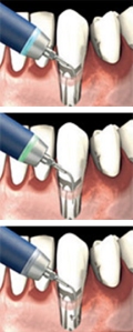
“PerioScan is an ultrasonic unit with treatment and diagnosis all-in-one, thereby offering new dimensions [sic] to periodontology. The unit detects and removes calculus by using a gentle method of treatment preventing the accumulation which causes periodontitis.
The tooth surfaces are being analysed by the touch of the ultrasonic tip (scaler) on the basis of the physical oscillation pattern. Indicator for the presence of calculus on the root surface is the blue LED lighting integrated into the tip of the handpiece. The LED colour changes to green when it contacts healthy surface. Two containers allow the use of different irrigating liquids during treatment.
The user interface is large and clear so that the user can have a glance at the data and the settings during the treatment at all times. The design impression and the colour concept of the interface are a reference for present and future projects of the entire product range of Sirona.”
The rationale for the devolpment of a smart device for both detecting (based on “fuzzy-pattern recognition”) and removing calculus (by conventional ultrasound) has certainly been recognition of a common disadvantage of any ultrasonic scaling device: its lack of tactile control of whether the entire subgingival root surface has actually been machined. I had used this device for years as a striking example for lack of clinical evidence after 20 years of development, apart from a 2008 pilot study (which actually tested validity of calculus assessment after extraction of the teeth), that it may actually lead to better results as regards the clinical response to subgingival scaling which might justify rather high acquisition costs. But there is more which should be used for teaching undergraduates.
